JC BITSOMANG EYE CLINIC — Healthy Eyes for a Brighter Life

Eye Disease Center

Backed by proven expertise, we safeguard your eye health with confidence

Corneal Transplant Surgical Training

Apollo Hospitals — a top 3 global hospital — located in Hyderabad, India.
Dr. Choi Gyeong-bae received corneal transplant training from Dr. Rajesh Fogla, the leading authority in corneal transplantation at Apollo Hospitals.

Director Choi — surgical training at Apollo Hospitals, India, supervised by Dr. Rajesh Fogla

(L) Dr. Rajesh Fogla and (R) Director Choi

[Director Choi, Corneal Transplant Surgical Training — Supervisor: Dr. Rajesh Fogla]

Corneal Transplantation

Anatomy diagram showing the position of the cornea

What is Corneal Transplantation?

The cornea is the transparent front window of the eye

When the cornea loses its transparency due to trauma, severe inflammation, or congenital causes — becoming opaque or clouded — light can no longer pass through, resulting in vision loss.

When such opacities cannot be treated with medication or laser, the damaged cornea must be removed and replaced with a clear donor cornea so that light can once again reach the inside of the eye.

Surgical Techniques

Illustration of lamellar (partial-thickness) keratoplasty

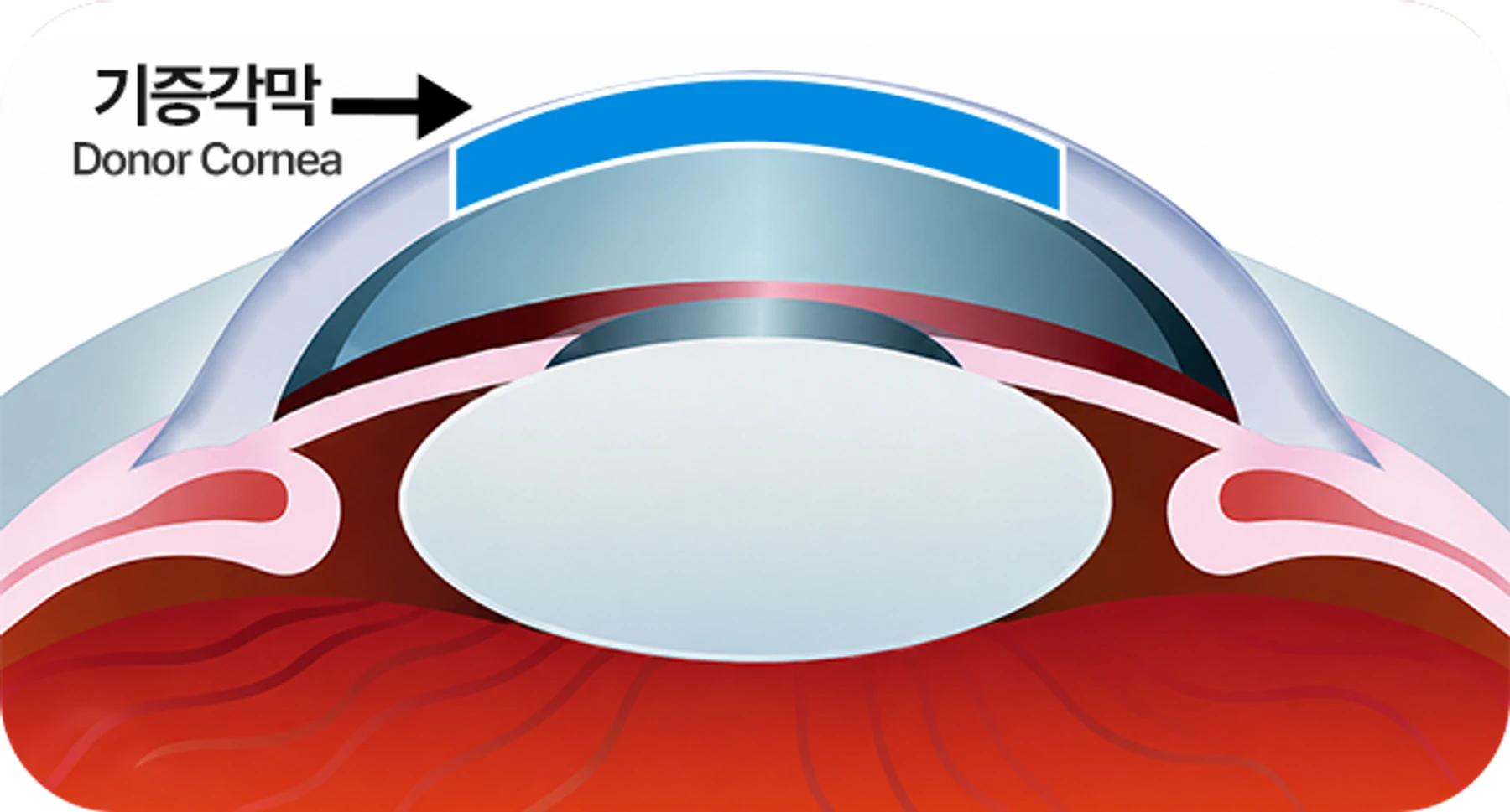
Lamellar Keratoplasty (DALK · DSAEK)

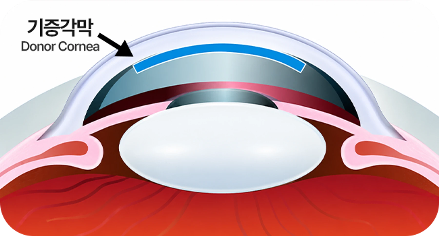
Lamellar keratoplasty replaces only the diseased layer of the cornea while preserving the patient's healthy tissue. When the corneal disease is limited to a specific layer — anterior or posterior — only that layer is removed and replaced with the corresponding layer from a donor cornea, reducing rejection risk and shortening recovery.

Illustration of penetrating (full-thickness) keratoplasty

Penetrating Keratoplasty (PK)

Most corneal disease involves the full thickness of the cornea. Penetrating keratoplasty makes a circular 6–8 mm incision at the central cornea and replaces it with a similarly sized clear, healthy donor cornea, which is then sutured into place.

Post-operative Care

  • Patients return for 5–6 follow-up visits within the first month, and continue scheduled examinations afterward according to the surgeon's instructions. Annual follow-up is also necessary to monitor long-term graft status.
  • An eye shield should be worn during sleep for the first month after surgery to prevent accidental rubbing of the eye.
  • Because the cornea heals slowly, sutures placed at the time of surgery are removed in stages over several months, while astigmatism is monitored and adjusted.
  • Avoid strenuous exercise and heavy physical labor for approximately one month after surgery and rest as needed.
  • Antibiotic and steroid eye drops are typically prescribed throughout the recovery period.
  • Possible complications include infection at the surgical site, rejection, glaucoma, astigmatism, and hyperopia. Most are treatable when addressed promptly, but delayed treatment can lead to graft failure.

Surgical Success Rate

One-year graft survival without complications or rejection ranges from 80% to 90%, with five-year survival of approximately 60–70% — a relatively high success rate. If the transplanted cornea later loses clarity due to complications or rejection, re-grafting with another donor cornea is possible.

Eye Donation

Eligibility for Eye Donation

Eyes from infants under two years old or adults over seventy are generally not used for corneal transplantation. Eyes from individuals with infectious diseases such as AIDS or hepatitis, or systemic malignancies such as leukemia, are also typically excluded.

Donation Procedure

Unlike other organs, corneas can only be donated after death. Donation can be registered in advance or arranged after death by family members, by contacting a corneal transplant registration center or the national organ transplant management center.

Eye Recovery and Preservation

Eye recovery should ideally take place within six hours after death. The eyelids are gently closed afterward, leaving the donor's appearance virtually unchanged. The cornea is then separated and preserved in a specialized 4 °C storage solution until surgery.

Transplant Waitlist and Donation Status

While both eye donations and corneal transplants are increasing, the number of donated corneas remains far below the waitlist demand. Greater public awareness of corneal donation is needed.