Retina & Glaucoma Center
Trusted expertise to safely protect your eye health.
Oral Medication
Oral medication serves as adjunctive therapy. The course varies by condition, but treatment typically continues for about six months.
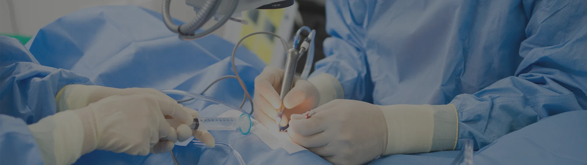
Intravitreal Injection
Ocular diseases — particularly retinal diseases — often cause reduced vision or partial visual field loss due to abnormal neovascularization. Vascular endothelial growth factor (VEGF) plays a key role in this process. Intravitreal anti-VEGF (antibody) injection and intravitreal triamcinolone (corticosteroid) therapy suppress endothelial proliferation and treat macular edema.
치료 전
Before Injection Treatment
치료 후
After Injection Treatment
The procedure begins with thorough disinfection around the eye followed by topical anesthesia. A fine needle is then used to inject the medication directly into the vitreous cavity. Intravitreal injection has long been performed and is regarded as a relatively safe procedure. By delivering medication directly into the eye, the required dosage and systemic side effects are minimized — making it an effective treatment option.
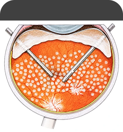
Panretinal Photocoagulation
After diabetes has persisted for more than a decade, diabetic retinopathy progresses from the non-proliferative stage to the proliferative stage. In the proliferative stage, abnormal neovascularization develops and intraocular hemorrhage from these fragile vessels can cause vision loss. Panretinal photocoagulation (PRP) is performed for proliferative diabetic retinopathy, and is now also applied in severe non-proliferative cases. During PRP, laser energy photocoagulates and destroys the ischemic (non-perfused) retinal areas where capillaries have been blocked. Treating these non-perfused regions halts new vessel formation and causes existing neovascularization to regress or disappear. This regression reduces the risk of vitreous hemorrhage and tractional retinal detachment — the leading causes of vision loss.
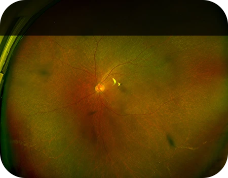
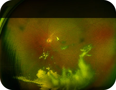
당뇨망막 출혈
Eye with Diabetic Retinal Hemorrhage
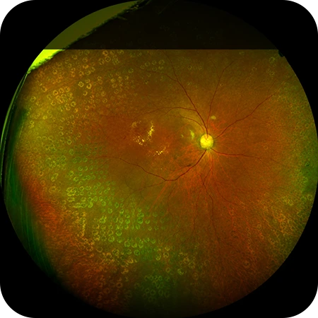
범망막 광응고술 PRP
Panretinal Photocoagulation
The procedure is performed mainly under topical anesthesia. After anesthesia, a specialized contact lens is placed on the eye to precisely focus the laser and prevent the patient from blinking during treatment. Once the lens is in place, a 532 nm wavelength laser is delivered to the retina.
Focal / Grid Laser Photocoagulation
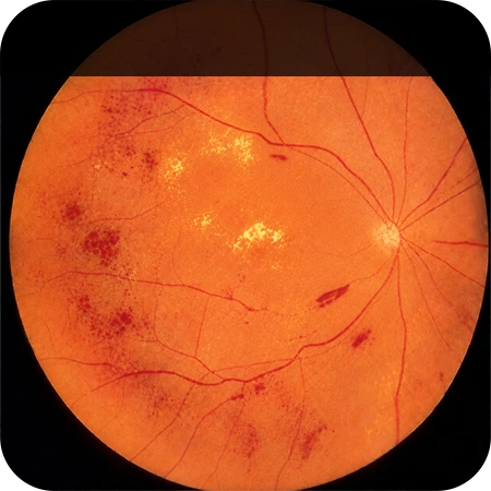
When retinal capillaries become occluded, the surrounding tissue forms new vessels to compensate. These neovascular vessels are fragile and rupture easily, causing hemorrhage and reduced vision. Laser photocoagulation is the appropriate treatment for this condition. After approximately three months, when the hemorrhage has partially resolved, fluorescein angiography is performed and retinal laser treatment is applied. Without laser treatment, retinal neovascularization may lead to vitreous hemorrhage and neovascular glaucoma — potentially resulting in blindness.
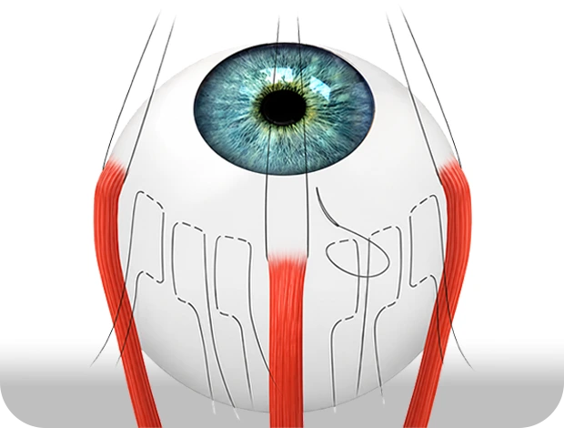
Scleral Buckling
Scleral buckling is a surgery that uses a silicone sponge or band placed on the outer surface of the eye to help the retina settle back into its original position. Once the retinal break closes, the subretinal fluid is fully absorbed and the detached retina reattaches to its original position. While scleral buckling can be performed under local anesthesia in the operating room, general anesthesia is frequently used due to the pain associated with the procedure.
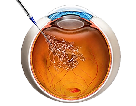
Pars Plana Vitrectomy
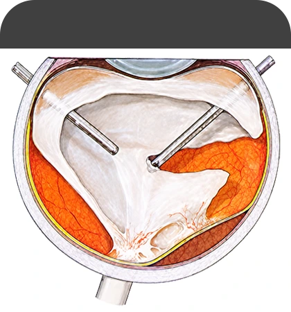
The vitreous is a transparent, egg-white-like fluid tissue that fills the eye, allowing light to pass through to the retina and helping maintain the shape of the eye. However, when hemorrhage, inflammation, or fibrous proliferation develops within the vitreous, opacification occurs and vision is impaired. In such cases, the vitreous must be removed to preserve or maintain the retina's intrinsic function against the risk of traction or detachment. Pars plana vitrectomy is a surgery that removes the intraocular vitreous using specialized instruments and reattaches the retina to its original state. After surgery, the patient must maintain a specific head position to support retinal recovery, and vision gradually recovers over several days to weeks as the intraocular air or gas is absorbed. Because retinal detachment can recur after surgery, postoperative hospitalization and regular follow-up examinations after discharge are essential to confirm retinal adhesion.
유리체 절제술
Vitrectomy
유리체 절제술 레이저
Endolaser Photocoagulation
Scleral Imbrication
Macular (retinal) detachment caused by macular hole most commonly occurs in high myopia. In high myopia, the axial length — the distance from the cornea to the retina — is longer than normal, which gives rise to such lesions. As a result, the retinal tissue weakens, leading to decreased central vision. In high-myopia patients, the axial length exceeds 26 mm (average axial length is 23–24 mm). Although growth should stop by age 20, the axial length continues to elongate abnormally, sometimes extending beyond 30 mm. Due to this effect, even with pars plana vitrectomy and intraocular tamponade (gas, silicone oil, or oil), the surgical instruments and tamponade fail to reach the retina, and in most cases reattachment is unsuccessful. Scleral imbrication is a surgery that shortens the elongated axial length by suturing the sclera in a specialized manner, and can maximize the effect of pars plana vitrectomy and intraocular tamponade surgery.