
Cataract & Presbyopia Center
Trusted expertise to safely care for your eye health.
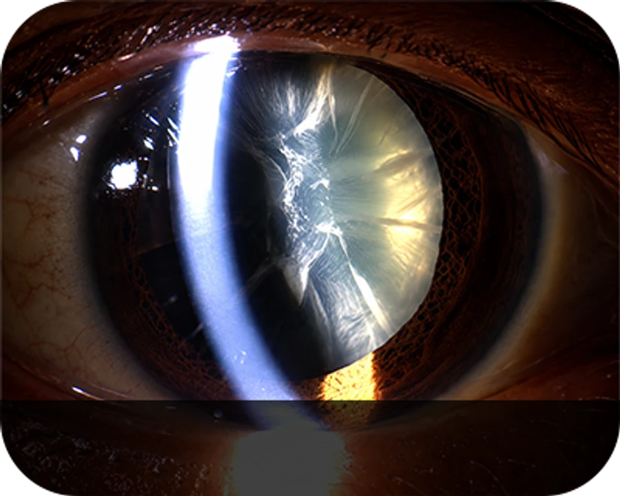
What is a Cataract?
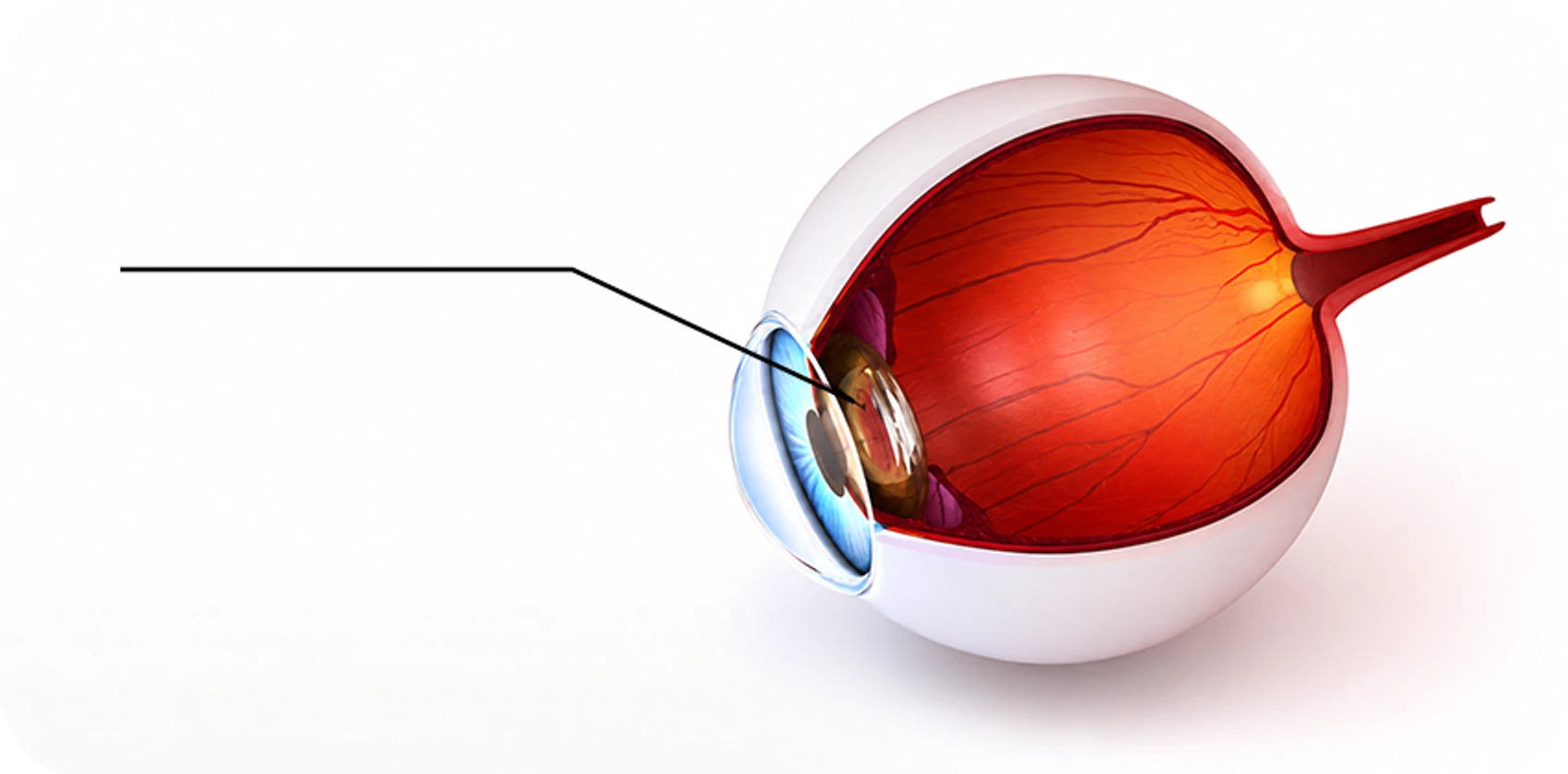
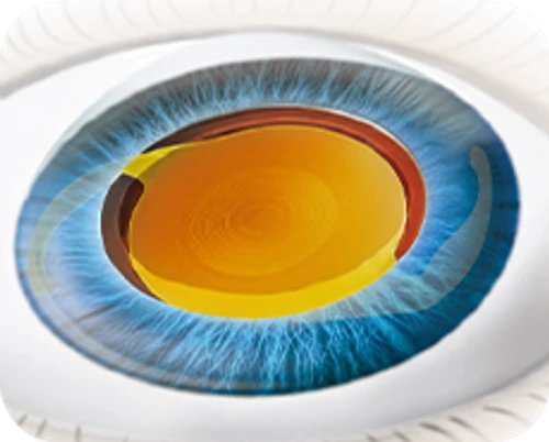
Cataract
A condition in which the lens becomes opacified, causing decreased vision
Cataract — opacification of the crystalline lens within the eye
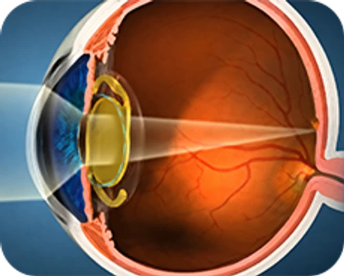
A cataract is a condition in which the normally transparent crystalline lens becomes opacified. The human eye is functionally and structurally analogous to a camera, with the crystalline lens corresponding to the camera lens. Just as a clouded camera lens yields blurred photographs, an opacified crystalline lens causes objects to appear hazy and produces progressive loss of visual acuity. Cataracts affect approximately 70% of individuals in their 60s, 90% of those in their 70s, and nearly all individuals aged 80 years and older.
 70%
70% 90%
90% 100%
100%Causes of Cataract
Age-related changes in the crystalline lens are the most common cause of cataracts. Other etiologies include hereditary factors, ionizing radiation and infrared exposure, steroid medications, psychotropic drug effects, and secondary cataract due to intraocular conditions such as uveitis; cataract may also progress after retinal detachment surgery, vitrectomy, or glaucoma surgery. In addition, congenital cataract — lens opacification present at birth — also exists.
- Congenital
Hereditary disorders,
chromosomal abnormalities, maternal drug exposure during pregnancy, congenital metabolic disorders. - Acquired
Senile (age-related) cataract,
diabetic cataract, traumatic cataract, atopic dermatitis, medications (steroids, antidepressants), intraocular disease (uveitis, etc.), UV-related ocular conditions.
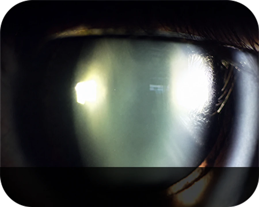
Cataract Symptoms
 시력저하Decreased Vision
Vision gradually declines, with a sensation that something is veiled over the eye.
 복시Diplopia
Objects appear doubled or tripled, overlapping in view.
 눈부심Glare
In dim or indoor lighting, light appears unusually bright or halated, making it hard to keep the eyes open.
 변색Discoloration
Whites take on a yellowish tint, and near vision may temporarily improve.
Cataract Surgery (Phacoemulsification)
* Depending on the post-operative ocular condition, transient intraocular pressure elevation, dry eye, or secondary procedures may be required. Please consult the specialist before proceeding.
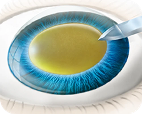 STEP 1
Incise the corneal region of the eye.
 STEP 2
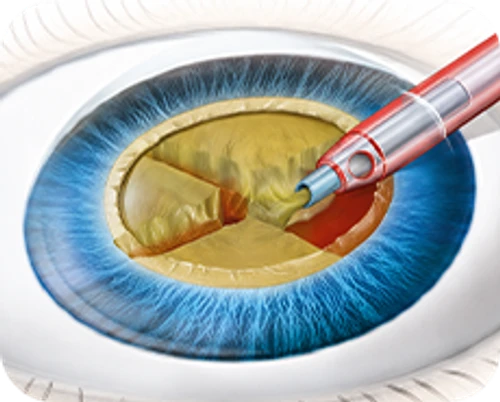
Using surgical instruments, create a circular anterior capsulorhexis on the lens.
 STEP 3
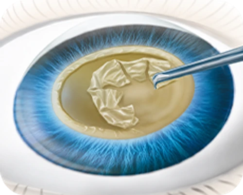
Fragment the opacified lens into small pieces.
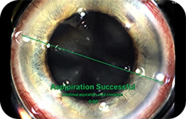
 STEP 4
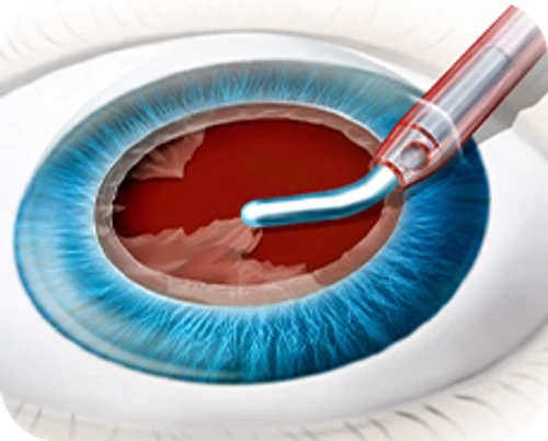
Aspirate and remove the fragmented lens material.
 STEP 5
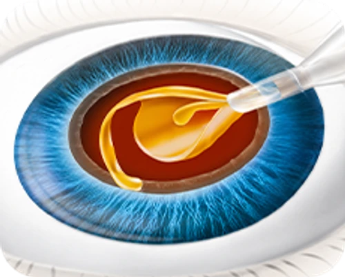
Insert an intraocular lens (IOL) into the empty capsular bag.
 STEP 6
Confirm that the post-operative focal point is precisely centered on the retina.
- STEP 1
Incise the corneal region of the eye.
- STEP 2
Using surgical instruments, create a circular anterior capsulorhexis on the lens.
- STEP 3
Fragment the opacified lens into small pieces.
- STEP 4
Aspirate and remove the fragmented lens material.
- STEP 5
Insert an intraocular lens (IOL) into the empty capsular bag.
- STEP 6
Confirm that the post-operative focal point is precisely centered on the retina.
Cataract Surgery (Laser)
 STEP 1
Use the laser to create the corneal incision.
 STEP 2
Use the laser to perform a circular anterior capsulotomy.
 STEP 3
Use the laser to fragment the opacified lens.
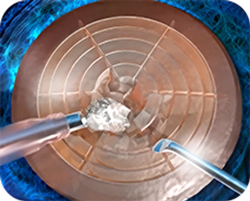 STEP 4
Aspirate and remove the fragmented lens material.
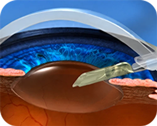 STEP 5
Insert an intraocular lens (IOL) into the empty capsular bag.
 STEP 6
Confirm that the post-operative focal point is precisely centered on the retina.
- STEP 1
Use the laser to create the corneal incision.
- STEP 2
Use the laser to perform a circular anterior capsulotomy.
- STEP 3
Use the laser to fragment the opacified lens.
- STEP 4
Aspirate and remove the fragmented lens material.
- STEP 5
Insert an intraocular lens (IOL) into the empty capsular bag.
- STEP 6
Confirm that the post-operative focal point is precisely centered on the retina.
What is Laser Cataract Surgery?
Premium surgery using a femtosecond laser
Laser cataract surgery employs precise laser incisions at the steps most prone to complications, followed by stable implantation of the intraocular lens (IOL). Unlike conventional surgery that depends on the surgeon's manual blade incisions, laser cataract surgery — performed with a femtosecond laser — creates corneal and capsular incisions according to precisely entered computer parameters, enabling a more accurate and safer procedure. Using ocular-navigation guidance, capsulorhexis and astigmatism correction are placed at the exact target location, combining cataract surgery with refractive correction in a more advanced surgical platform. The femtosecond laser also performs lens fragmentation, improving stability and accuracy and resulting in faster recovery and more satisfactory visual outcomes.
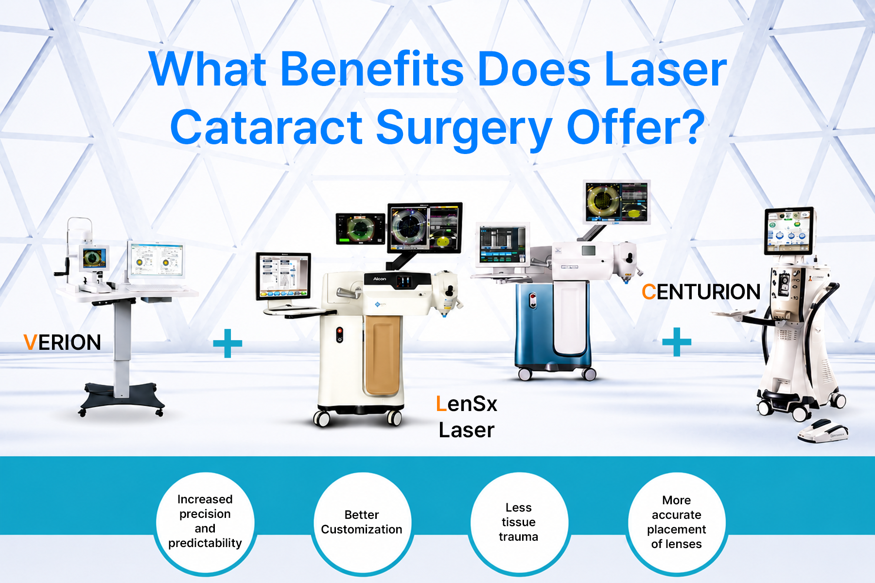
Advantages of Laser Cataract Surgery
Laser-precise incisions without error
The cornea is incised at the planned location with exact size and depth, and a perfectly circular capsulorhexis is created with the precise size and shape required, minimizing post-operative residual refractive error that could reduce target visual acuity — delivering a predictable visual outcome for the patient.
Astigmatism correction via limbal relaxing incisions
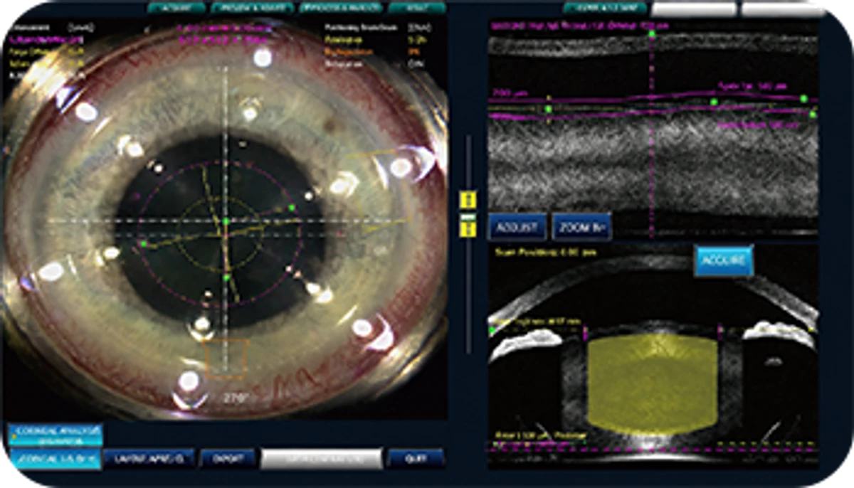
Limbal relaxing incisions (LRIs) can be placed at the exact location, size, and depth required. Corneal topography and wavefront aberrometry guide intraoperative measurement of corneal curvature radius for precise incisions, significantly enhancing the effectiveness of astigmatism correction.
Reduced complications from laser energy
Because corneal incisions, capsulorhexis, and lens fragmentation are all performed by laser, complications associated with manual surgery are reduced and visual recovery is faster.
Greater patient comfort
With laser-assisted cataract surgery, the incidence of corneal abrasion or epithelial defect, pain, infection, bleeding, intraocular structural damage, aqueous leakage, and anterior chamber collapse is markedly reduced, allowing patients to undergo cataract surgery with greater comfort.
* Depending on the post-operative ocular condition, transient IOP elevation, dry eye, or secondary procedures may be required. Please consult the specialist before proceeding.
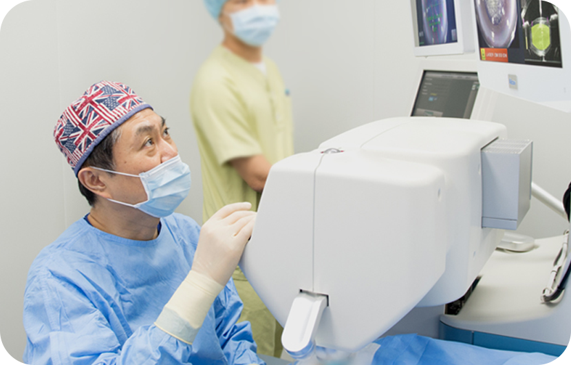
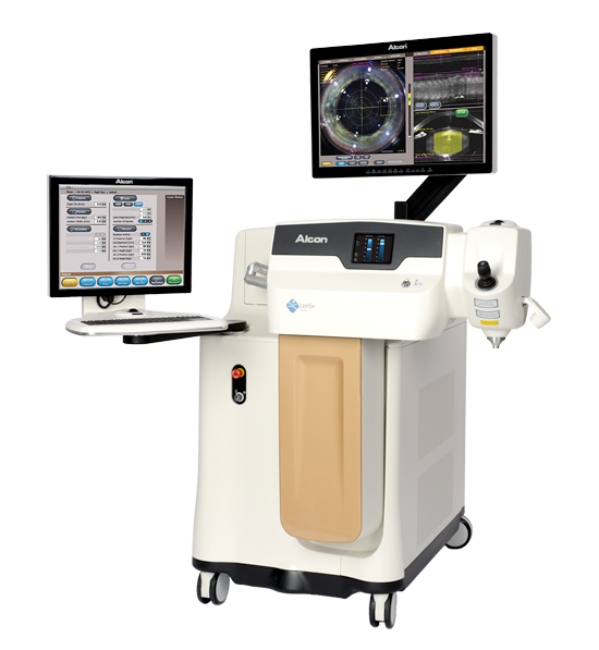
LenSx®
Precise laser incisions and lens fragmentation, plus corneal incisions for accurate laser-guided astigmatism correction.
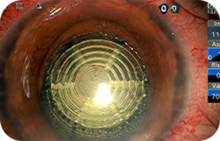
Conventional cataract surgery has long relied on blades and ultrasound — blades to incise the corneal tissue and lens capsule, and ultrasound to remove the cataractous portion of the crystalline lens. However, in the hands of less-experienced surgeons, blade-induced irregular astigmatism, thermal damage at the incision site from ultrasound, or corneal endothelial cell loss from ultrasound waves could lead to slower recovery or post-operative astigmatism. These limitations have been almost entirely addressed by a new-generation premium cataract surgery platform: the LenSx® femtosecond laser cataract surgery.
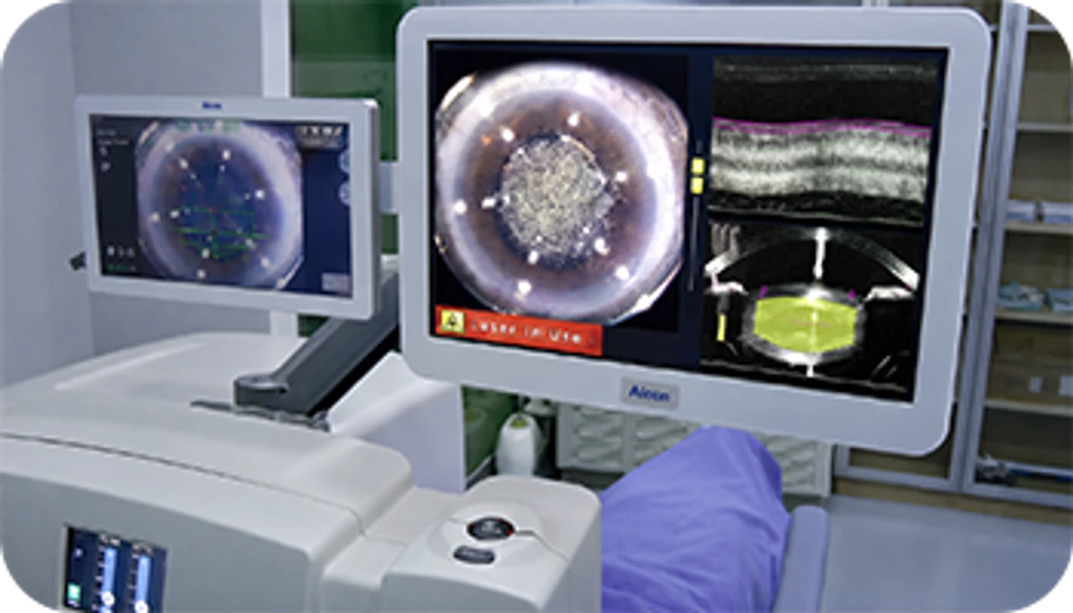
LenSx® uses ocular-navigation guidance to perform precise capsulorhexis and astigmatic correction at the exact target location, combining cataract surgery with refractive correction in a more advanced surgical platform. The laser also performs lens fragmentation, further improving stability and accuracy and resulting in faster patient recovery and more satisfactory surgical outcomes.
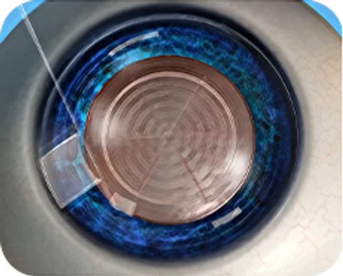
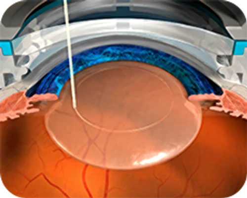
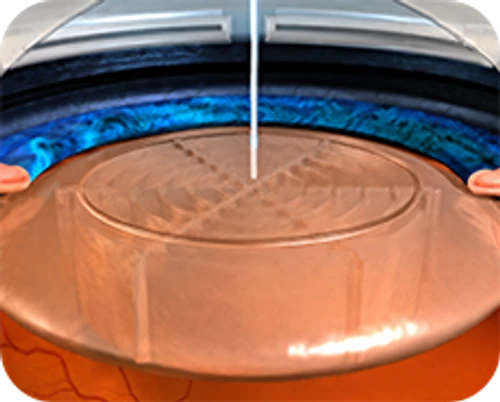
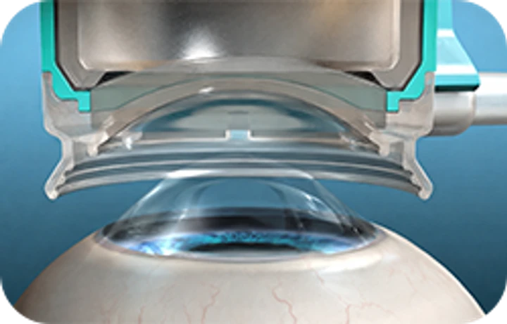
![LenSx cataract surgery step-by-step diagram — [Laser Anterior Capsulotomy]](/static/cataract/about/sections/lensx-proc-1.webp) [Laser Anterior Capsulotomy]
[Laser Anterior Capsulotomy]![LenSx cataract surgery step-by-step diagram — [Laser Cataract Fragmentation]](/static/cataract/about/sections/lensx-proc-2.webp) [Laser Cataract Fragmentation]
[Laser Cataract Fragmentation]![LenSx cataract surgery step-by-step diagram — [Laser Corneal Incision Creation]](/static/cataract/about/sections/lensx-proc-3.webp) [Laser Corneal Incision Creation]
[Laser Corneal Incision Creation]
Benefits of LenSx Laser Cataract Surgery
- Safety
The most demanding steps of cataract surgery — capsulorhexis, lens disassembly and fragmentation, astigmatism correction, and primary incision — are performed automatically by the LenSx laser, enabling a safer procedure.
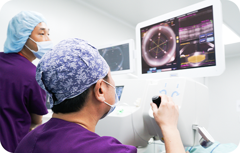
- Precision
OCT (optical coherence tomography) measures the eye's internal structure, and a computer plans and designs the procedure before it begins. Incision range and position can be set to within one-thousandth of a millimeter, delivering precision and accuracy beyond any other method. In particular, capsulorhexis, primary incision, and astigmatism correction directly influence post-operative refractive outcome, so the laser-driven precision ensures the planned refractive result is achieved.
- Speed and comfort
Femtosecond (one-millionth of a second) laser pulses make the procedure extremely brief. Because the crystalline lens is pre-fragmented by laser before surgery begins, ultrasound phacoemulsification time is shortened, allowing both patient and surgeon to proceed in greater comfort. With less ultrasound energy applied to the eye and a shorter operative time, faster post-operative recovery can be expected.
- Suitable for difficult cataracts
When the cataract is severely advanced or the eye's anatomy is challenging — shallow anterior chamber, partial lens subluxation, weak zonules — conventional surgical techniques may not be feasible. Even in such difficult cases, LenSx can perform incision and lens disassembly without applying force to the crystalline lens, allowing surgery for high-risk cataracts to be completed comparatively easily and safely.
LenSx Cataract Surgery Video


