
About JC Bitsomang Eye Clinic
Always by your side, listening to every patient — that is the JC Bitsomang Eye Clinic we strive to be.
Always by your side, listening to every patient — that is the JC Bitsomang Eye Clinic we strive to be.
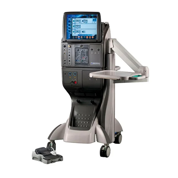
Vitreoretinal Surgery System
Alcon Constellation Vision System integrates intra-vitreal gas injection, intraocular laser, and 10,000 cpm ultra-high-speed vitrectomy in a single platform — used for same-day retinal surgery as well as complex cataract cases at risk of complications. Designed to minimize vision loss due to surgical complications.
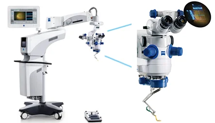
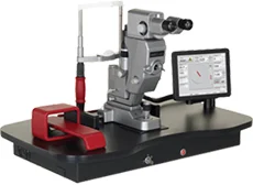
Surgical Microscope with Integrated OCT
Carl Zeiss RESCAN 700 supports corneal transplant, retinal, cataract, and astigmatism procedures with real-time 3D intraoperative OCT visualization. Surgeons can confirm intraocular details in real time for safer, more confident decisions.
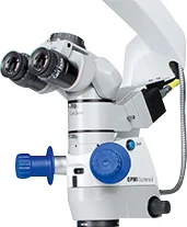
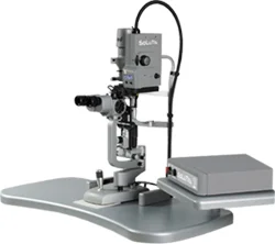
Surgical Microscope
Built on world-renowned Carl Zeiss optics with Stereo Coaxial Illumination (SCI™) for an outstanding view quality, an auto-adjusting Depth of Focus system, and a Retinal Protection Device — maximizing surgeon convenience and patient safety.

OCT Angiography
Captures the retina, choroid, and RPE for tracking comprehensive retinal disease — dye-free imaging, twice the previous speed, high-resolution mode, and eye-tracking deliver convenience and precision matching university hospitals. Highly effective for diabetic retinopathy and macular degeneration.
Fluorescein Angiography
Specialized for central retinal imaging and choroidal vasculature with 30°/55° lenses plus an Ultra-Widefield lens for fine detail. Performs simultaneous ICG and FAG in a single shot and supports video capture, ideal for tracking fluorescein flow from onset — particularly effective for AMD diagnosis.
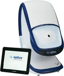
Fluorescein Angiography
Non-mydriatic imaging covering 80% of the retina (200°) for fast and comfortable pre-surgical retinal evaluation. Supports basic fundus, autofluorescence, fluorescein angiography, and ICG angiography modes.
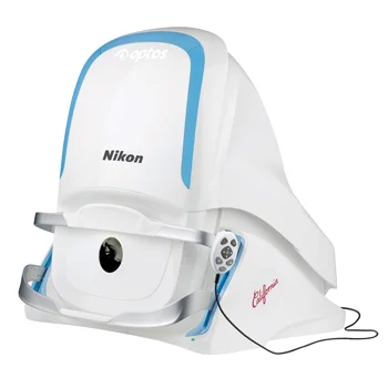
Non-mydriatic Ultra-wide Fundus Camera - 2 units
Optomap Daytona captures 80% of the retina (200° field) without pupil dilation — enabling fast and comfortable pre-surgery retinal screening.
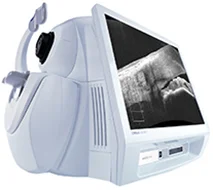
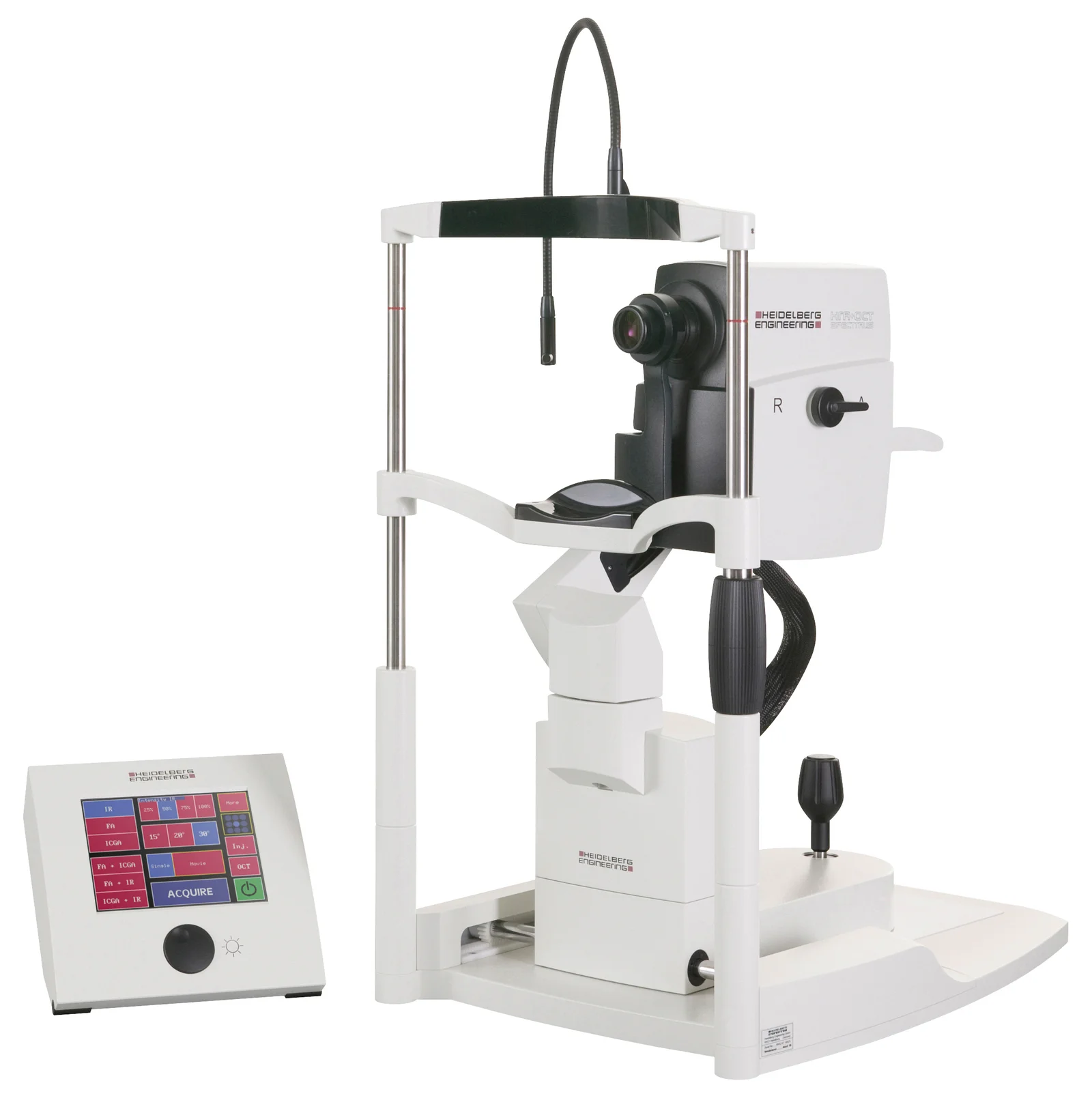
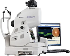
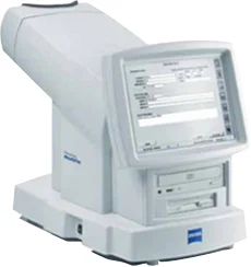
Optical Coherence Tomography
Carl Zeiss CIRRUS HD OCT-5000 is the latest ophthalmic OCT, used by select major university hospitals in Korea. Essential for early diagnosis across all eye diseases — especially retina and glaucoma — it uses spectral characteristics of light for faster imaging at higher resolution. Uniquely enables dye-free fluorescein-style fundus imaging, eliminating contrast agent side effects and providing patients with comfortable, hassle-free exams.
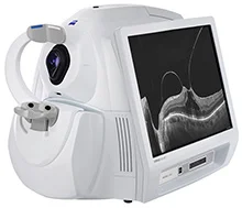
Optical Coherence Tomography
Inspects optic nerve and retina with 5-micron precision (1/2,000 of 1cm). Provides 3D high-resolution retinal diagnosis and excels at optic nerve thickness/pattern analysis — specifically designed to detect early-glaucoma nerve fiber thinning and damage.
3D Optical Coherence Tomography
3D OCT for the optic nerve and retina, enabling whole-eye precision exams that were previously impossible. Built-in FAG further improves accuracy. · OCT + COLOR + RED GREEN + FAG + ANTERIOR · Over 10 megapixel fundus imaging.
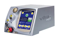
Photodynamic Therapy Laser
Closes choroidal neovascularization in macular degeneration. Unlike conventional thermal lasers that also damage normal tissue, PDT uses a photosensitizer and low-energy non-thermal light to selectively close pathological neovessels.
Glaucoma Treatment Laser
SLT (Selective Laser Trabeculoplasty) is a modern glaucoma treatment. Compared with traditional ALT, SLT safely lowers IOP without tissue damage or side effects by targeting only melanin-pigmented cells, leaving the trabecular meshwork untouched.
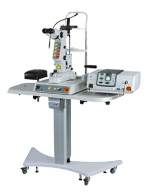
Retinal Therapy Laser
The PASCAL retinal laser allows treatments previously divided into 3–4 sessions to be completed in one, with reduced patient discomfort. Macular edema — a common side effect that can reduce vision — is also minimized, making it very safe. The same platform supports SLT (Selective Laser Trabeculoplasty) for glaucoma.
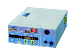
532nm Laser
NIDEK GYC1000 is a CW LBO-based green argon laser for intraocular treatments during vitrectomy (diabetic retinopathy, retinal tears, retinal detachment, etc.), delivering 1700mW of corneal-incident power — the highest available in Korea.
532nm Laser
Proven across diabetic retinopathy, retinal hemorrhage, and detachment, as well as glaucoma treatment and prevention. The advanced Hi-Valuable Crystal Cavity (upgraded from liquid cavity) produces a more stable 531nm beam, while built-in safety filters ensure maximum reliability in any situation.
Automated Perimetry
Early-glaucoma-detection perimeter with superior sensitivity vs. legacy devices, and rapid result generation that minimizes total exam time.
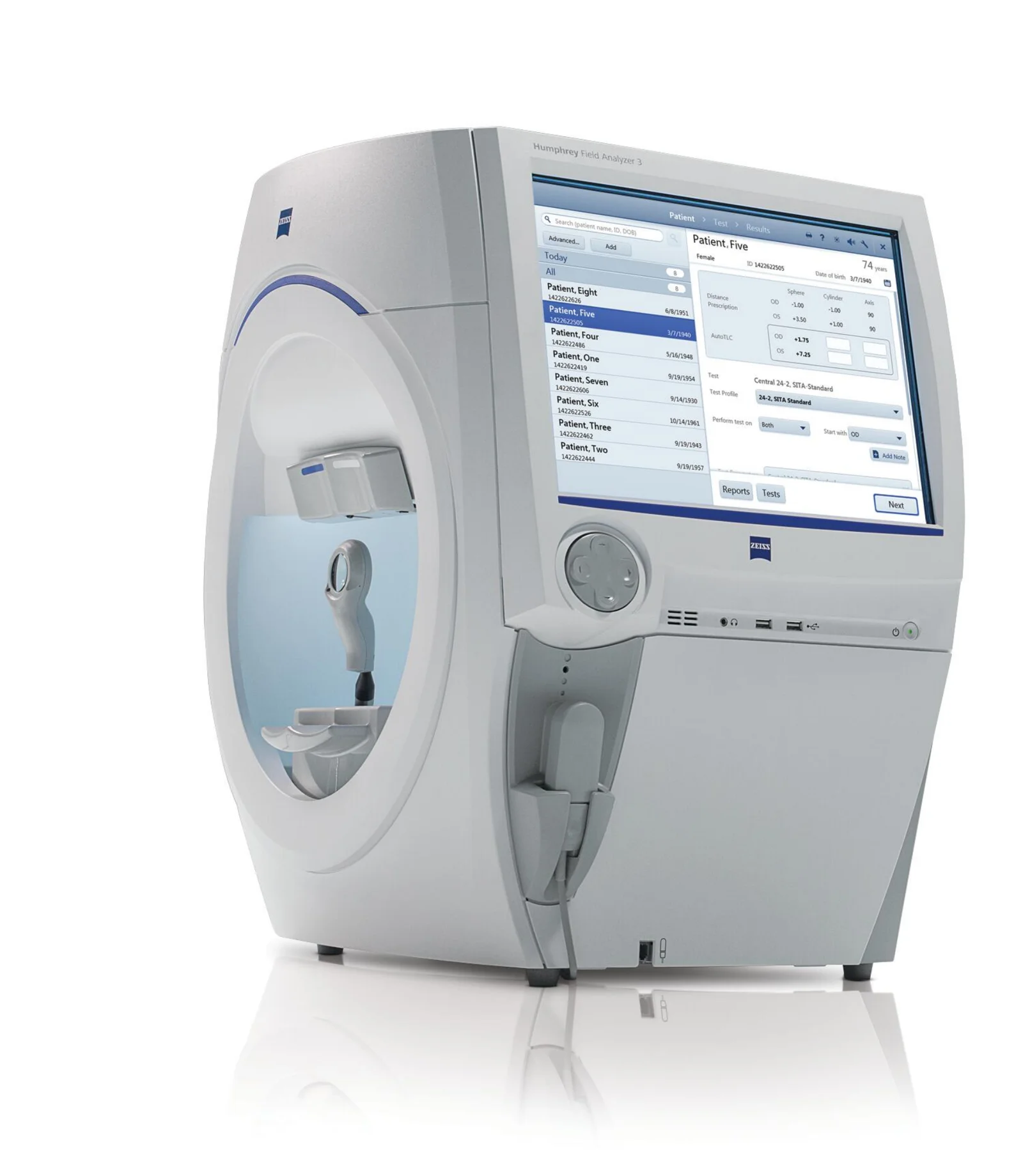
Automated Perimetry
HFA3 840i is the latest generation of the widely used Humphrey perimeter, enabling precise visual field exams and glaucoma progression analysis. SITA Faster shortens exam time without sacrificing accuracy, and STATPAC enables systematic comparison/analysis of visual field changes over time. Blue-on-Yellow perimetry detects early glaucomatous changes with greater sensitivity, and Gaze/Head Tracking further improves exam reliability.
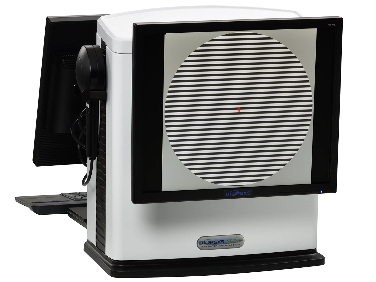
Electrophysiology Testing
Diopsys ERG electrophysiologically evaluates retinal and optic nerve function — ERG (electroretinography) and VEP (visual evoked potential) measure electrical responses of visual cells. Used to diagnose functional retinal/optic-nerve abnormalities and to evaluate retinal function before and after cataract surgery.
Precision Fundus Camera
A fundus camera that photographs the retina to diagnose intraocular abnormalities — essential for diagnosing and monitoring diabetic retinopathy, macular degeneration, and glaucoma.


